The Advantages of Laser Gum Surgery
The Phase Contrast Microscope
In 2009 I figured out a unique way to use a phase contrast microscope and transfer the video image live onto a high definition TV screen. A microscope expert showed me how to port the image through a DSLR to a HD TV screen for live culture presentations in real time for the patients to see.
When Covid hit, I stopped using the microscope. However, I’ve started using it again because I now have a periodontist on staff. The microscope is such a great visual aid, because it shows us the microbiome in our patient’s mouths. You have heard of the saying, ‘A picture is worth a thousand words.’ A video is worth even more.
Combining HD video with a microscope has allowed me to show patients an underlying cause of their inflammatory condition that is responsible, in part, for their persistent and worsening periodontal condition. It’s very powerful imagery that puts into perspective what’s causing gum disease.
Combining Technologies in Unique Ways
We have incorporated the microscope into our perio treatment in conjunction with LANAP laser gum surgery.
Combining the following technologies to give patients results they cannot achieve any other way. These results include faster healing times, less pain and trauma, and being able to combine multiple periodontal surgical techniques in one surgery. Here are the modalities Dr O and Dr Bae use to heal our patient’s gums:
- The Millenium laser
- The Fotona Nd:YAG laser
- The Phase Contrast Microscope before and after
Case Study
Recently we had a lovely, health-minded patient fly in from Denver to treat her gum disease, a referral from another dentist who is aware of our work with the microscope. Her hygiene was impeccable, but her case was still active and getting worse, to the point of losing teeth.
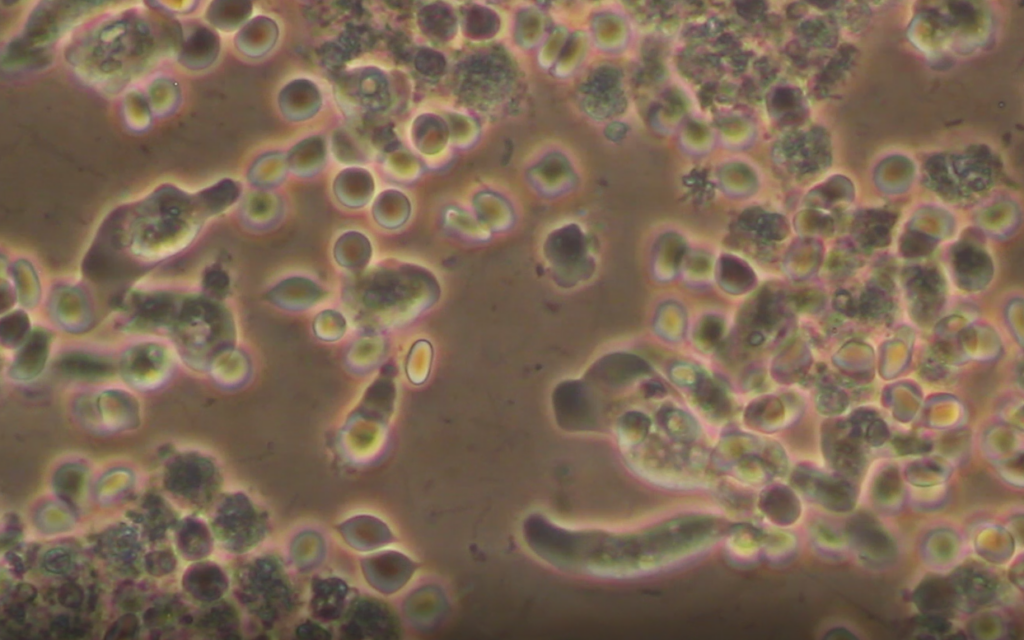
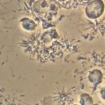
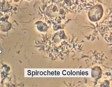
Her microscope video revealed parasites and large immune cells to try to fight them. When it comes to advanced microbes like amoeba, the immune cells always fail. The patient’s question to us was, ‘What am I missing; what am I doing wrong?’
We did a microscope evaluation beforehand and identified the microbes and amoeba that were keeping her from healing or getting better with any other treatment.
Once she could clearly see the reason why all her previous efforts had failed, she was very anxious to pursue the laser approach at our office.
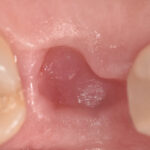
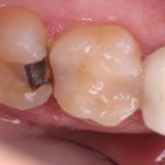
X-rays from the day of laser surgery
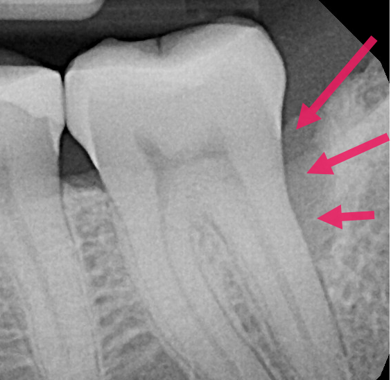
Here is a before x ray on the day of the surgery showing one of the boney defects with the red arrows. You can see by the dark defect that the patient had lost a lot of bone behind the back tooth. This essentially is a breading ground for bacteria and parasites to infect the tooth and cause more bone loss and she would have eventually lost the tooth.
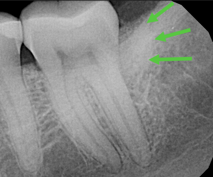
Here is the after x-ray showing the boney defect grafted. You can see the defect in the bone has been repaired back up to it’s original and healthy level. See green arrows and the normal look to the bone behind the back tooth instead of the black area before.
By using multiple modalities and technologies, not only were we able to treat someone from out of state with advanced periodontal surgery, we were able to send her home in only one week. Also, her post operative swelling and discomfort was only slight to moderate, and she reported that she did not need any pain medication.
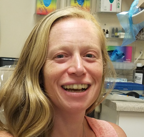
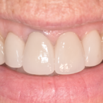
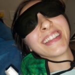
Here is Erin smiling 3 days after surgery, with minimal pain or swelling. When does that happen after gum surgery? The short answer is, almost never!
Carey O’Rielly DDS has been a practicing dentist for 35 years. He went to USC Dental School and Duke University for his undergraduate degree. He grew up in Laguna Beach and now lives in La Costa with his wife Victoria, who runs his office.
He began his career by owning and operating a network of six offices in the San Francisco Bay Area. Presently he owns a private holistic practice in North County San Diego’s Encinitas.
Dr. O started looking for solutions to his health challenges that resulted from the stress and environmental toxicity that built up over a ten year period running his dental network. He has dedicated himself to learning about oral systemic problems and how dentistry can affect your health. He has applied what he has learned over the last twenty years to ensure he, his staff and his patients are protected from the chemicals and toxic materials found in most dental offices. He has produced an environmentally friendly office that is also peaceful and calm.
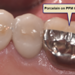
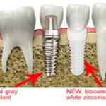
He is an expert on dental materials having looked at hundreds of biocompatibility lab tests over the years. He has identified the most bio-friendly materials to use in his practice and which dental materials can be used to replace metal fillings and crowns, including BPA free and fluoride free ‘white’ fillings. He also uses metal-free Zirconia or ceramic implants and PRF (platelet-rich fibrin) grafting materials which come from the patient’s own blood.
Dr. O’Rielly teaches C.E. courses on the systemic effects of gum disease. He is an expert in using phase contrast microscopy for analyzing dental infections, where he shows patients what kind of microbes, i.e. bacteria, amoeba, and yeasts like candida are populating the mouth and affecting the body as a whole.
He has an educational blog and is writing a book on dental health called ‘Hidden Dental Infections: Healing Root Canals and Infected Teeth with the Erbium Laser’ where he discusses dental nutrition, toxic dental materials and the effects of old root canals on inflammation and overall health.